Have a suggestion to improve this page?
To leave a general comment about our Web site, please click here
Imagine the Unimaginable Harnessing the Power of DNA: Principles of Genetic Engineering
byLaura Carroll- KochIntroduction
Imagine a band- aid that when applied fuses and heals the skin or a bio-lens that when placed on the eye corrects one's vision; or cancers, when discovered, can be attacked and destroyed by nano-armies, armed with healing genetic arsenals, DNA warriors. This is the potential of the science of genetic engineering.
We live in an exciting age, a New Age Renaissance of science and technology, harnessing the powers of DNA to transform medicine; the diagnosis, treatment and prevention of disease. The forward thinking cultures of biology, medicine, genetics, and engineering are exploding with new information and insights, creating highly active, dense networks of shared ideas. Cutting-edge technology is revolutionizing how we are able study, find, and correct genes within DNA. Genetic engineers are now exploring unchartered territories of the human genome, resulting in groundbreaking discoveries and treatments, offering hope to the once hopeless.
The advancements in technology are unraveling our DNA with lightning speed, decoding the human genome and revealing its long held secrets. As a result, we have the ability to manipulate the human gene and harness the power of DNA. The secrets that lie within its codes are now available, the way we use this knowledge is in the hands of the next generation, our students. Its outcome limited only by one's imagination. So, let us begin, let us ask our students to imagine the unimaginable.
Consequently, this calls for a shift in the direction of education, a profession devoted to the advancement of learning and the acquisition of knowledge. Can we offer our students a glimpse of the future, a vision of possibilities, and the language to discuss it? Should these concepts be in our classrooms now, infused into the curriculum, improving what students know and are able to imagine for the future? I believe they do. This unit will offer needed insight for future opportunities in bioengineering by growing a culture for students to develop their abilities to think, speak, and explore its concepts. It is an important shift. Our students are capable of understanding these concepts and learning the language of DNA; if given the opportunity. Ultimately, we want our students to be academically prepared for potential fields of science, ready to make their own mark, participating and contributing to the innovative solutions of our era, a new generation excited and prepared to tackle the challenges before us.
In an effort to meet these academic challenges, this unit will create a forum for students to learn the fundamental concepts of genetic engineering and the acquisition of its language. Through a study of the human cell, its structure, and function, students will learn the elements of cell biology. An introduction to the structure and function of DNA and the basic processes of replication, translation, and transcription will afford students the conceptual background to understand the methods in which genes can be modified. Insight into the manner in which genetic material is passed through generations will be discovered when students trace genetically inherited traits through their families. Genetic therapies will be explored in relation to specific illnesses as a way of learning the steps needed to create individual genetic treatments. Within this context, students will learn the ways in which DNA can be altered and delivered into a cell; thus providing a reservoir of knowledge to create a personal engineering toolbox of vectors and therapies for the repair and treatment of genetic disorders. In the end, students will apply these concepts in a project where they create their own gene therapy to correct a genetic illness. Ultimately, the purpose of this unit is to offer students a glimpse into the world of genetic engineering, inspiring a new generation prepared, excited, and empowered to advance the landscape of global health.
Rationale
I teach fourth grade at John S. Martinez Elementary School in New Haven, Connecticut. My class is comprised of approximately twenty-seven students who are 98% Hispanic. John S. Martinez is in an urban setting where the students are from low income families. Within the classroom, my students are strapped with the demands of testing: CMTs, (Connecticut Mastery Test) as well as district math, reading and language arts assessments. Reading, writing, mathematics, social studies, and science compete for center stage, often squeezing the science curriculum to its bare bones. Students are unprepared for the rigors and expectations of the science CMT's in fifth grade. We are living in an age of exploration and enlightenment where science and technology offer a plethora of information accessible to our students. I would like to bring science to center stage, wrapping the curriculum around it, making it the star our students need it to be; shining its light across the curriculum through the engaging, intriguing subject matter of the cell, DNA, genes and the science of bioengineering .
My goal is to stimulate excitement in biological science by focusing on the properties of DNA and its functions within the cell. Since my students do not have background knowledge in cell biology, this unit will begin with an introduction to cell structure, and function. Then, the fundamental principles of DNA will be introduced through the study of its characteristics, structural elements, and functions within the human cell. Through this exploration, I expect students to form an understanding for the scope and magnitude held within our DNA. I hope that pure awe and wonder will evolve as students come to realize that DNA is the building block of life, holding the design of who we are and ultimately who we become. A focus on the role of genes in the inheritance of traits and diseases will be exercised with the Punnett square. In addition, students will learn that the genes in our DNA hold our individualized human blueprint in each one of our cells. Next, students will learn that it is through the expression of the genes in each cell, the way they turn off and on, that defines cell function. Then, students will learn that the processes of DNA replication, transcription, and translation ultimately determine the proteins built which make our cells function properly or produce disease. Finally, students will study genetic illnesses, their causes, symptoms and the ways in which gene therapies, interventions, or engineering to correct these illnesses can be applied. A culminating activity will be for students to imagine the unimaginable, applying what they learned to create their own DNA therapy for an illness.
I envision an interdisciplinary unit of study involving mathematics, science, reading, and writing. Concrete activities and visual illustrations will help to teach complicated, abstract ideas. After students learn concepts, they will have opportunities to demonstrate this knowledge in a variety of ways, such as models, simulations, stories, songs, poems, visual descriptions, and written explanations. In addition, students will keep a daily journal, recording concepts, procedures, and questions as well as documenting learning through illustrations. Students will cooperatively reflect upon learning daily and formulate new questions in their journals.
A revolutionary age of science and technology is before us, where what was once a dream is now a reality. Genes are now used as tools to treat, cure, and prevent disease. Imagine what our students will be able to develop if given this knowledge to ponder, simmer, and grow; empowered to envision future treatments of their own. I cannot think of a more relevant topic than genetic engineering at this time. With this knowledge, I believe I can inspire my students to think, question, investigate, and imagine ways in which they can change the world, improving the quality of life and health on our planet.
The Human Cell: Anatomy and Function
The cell is the microscopic unit of all living organisms. We have 50,000,000,000,000 (50 trillion) cells in our body. Through its evolution, the human cell has become a highly developed, complex structure of organized systems (see Figure 1). This cell is able to reproduce, respond to stimuli, maintain homeostasis, grow, and adapt to the environment. 1 A double bilayer membrane surrounds the cell like a wall. This membrane enables the cell to regulate the flow of water and materials through this membrane. The cell is filled with a fluid called cytosol allowing the organelles to float. In addition to the organelles, many proteins float in the cytosol controlling cell metabolism. Cytoplasm is the term used to describe the cytosol and all the organelles floating within it. Each organelle is encased in its own membrane, has a unique structure and a specific function. 2

http://en.wikipedia.org/wiki/Cell_%28biology%29
Figure 1 A Human Cell
Schematic diagram of the human cell illustrating the structure and position of the organelles within the cell structure.
Organelles
The organelles are the parts of a cell. They include the nucleus, the nucleolus, ribosomes, smooth endoplasmic reticulum, rough endoplasmic reticulum, mitochondria, golgi apparatus, lysosomes, centrosome, and peroxisomes. Our DNA (deoxyribonucleic acid) is located in the cell nucleus, the largest organelle (see Figure 1). Like the cell membrane, the nuclear membrane is a porous bilayer of lipids regulating the flow of material into and out of the nucleus. The DNA controls every cell process and the ways in which our cells communicate and work together creating our unique individual design. The same DNA is within every cell in our body. The nucleolus sits within the nucleus of the cell making ribosomes and storing RNA. Ribosomes are stations where protein synthesis occurs. The smooth endoplasmic reticulum (ER) has a smooth surface and is an extension of the nuclear membrane. Rough endoplasmic reticulum looks bumpy because of the many ribosomes sitting on its surface synthesizing proteins. The proteins are also stored in the rough endoplasmic reticulum before being transported to the places where they are needed in the cell. The mitochondria are the energy sources of the cell producing and storing the energy for all the cell processes and movement including division and replication. Similar to the nucleus, the mitochondrion is encased in a bilayer membrane. The inside of the mitochondrion is a maze of folds called, cristae, increasing the inner membrane surface layer. ATP, the primary source of cell energy, is produced on this surface area when sugars are combined with oxygen. The golgi apparatus packages molecules so they can be transported to other parts of the cell. Lysosomes are little organelles that have digestive enzymes within them that digest unwanted particles in the cytoplasm. A centrosome is an organelle that contains a small pair of organelles called centrioles. During cell division, the centrioles replicate and the centrosomes divide, moving to each end of the cell nucleus. At each end of the cell, the centrioles make a spindle in the process of cell division. Peroxisomes are organelles that detoxify the cytosol by breaking down the hydrogen peroxide into water and oxygen. 3 Another way to think of the cell and its organelles is to compare it to a school.
A Cell Is Like A School

qcpages.qc.cuny.edu/~esornberger100/Worksheet.doc
DNA Structure

Figure 2 Structure of the DNA Molecule
The DNA molecule is made of two polynucleotide strands spiraled around each other to form a double helix. The polynucleotide strands are composed of a five-carbon sugar molecule, phosphate group, and nitrogenous bases. (thymine, adenine, cytosine, guanine)The strands are connected by nitrogenous base pairs (A-T and C-G) which are held together by a hydrogen bond. The bases adenine and thymine will always pair and the bases guanine and cytosine will always pair on the DNA molecule.
DNA, or deoxyribonucleic acid, is a large molecule that holds the genetic information of our entire human design, a blueprint of the human form and function; the plan for whom we are and what we are likely to become. DNA is encoded with the instructions for the construction of all our physical structures and the plethora of their functions. A spool-like protein, called histone, bundles this massive amount of DNA in extremely condensed, tightly packed chromosomes that enable it to fit within the nucleus of the cell. If you lined up the entire DNA from all the cells in your body, its length would be 6,000 million miles long! 4
The Nucleotide

Figure 3 A Nucleotide
A nucleotide is a DNA unit composed of a five-carbon sugar, a phosphate group, and a nitrogenous base. The sequence of these units forms the DNA molecule.
The structure of DNA was established by James Watson and Francis Crick and published in the journal Nature in 1953. This work determined that DNA is arranged in a double helix, meaning it is composed of two polynucleotide strands in a ladder-shaped spiral. When untwisted, the DNA spiral can be compared to a ladder. (see Figure 2) The "rails" of the ladder, or the DNA backbone, are made of phosphates and sugar molecules. The "rungs" of the ladder are made of four different nitrogenous bases. A nucleotide is a unit composed of a five-carbon sugar, a phosphate group, and a nitrogenous base.(see Figure 3) Each base is attached to the sugar molecule part of the "rail." These four chemical bases are the building blocks of the DNA molecule. They are A (adenine), T (thymine), G (guanine), and C (cytosine). Each base is paired with its partner base on the DNA molecule by a hydrogen bond. Adenine is always paired with thymine and guanine is always paired with cytosine (i.e., A-T and C-G). Consequently, these pairings are called base pairs in DNA language. These base pairs are the "rungs" on the DNA ladder that join the two rails or two DNA strands. There are about three billion nucleotides, or base pairs, in the human genome in every cell. If the ladder was climbed, from one end of the DNA molecule to the other, the rungs on the DNA spiral-ladder could be read in order: this sequence (or order) is the language of DNA. The unique sequencing of the bases is the way DNA encodes the information it carries. Further, since these four bases will always pair with their complimentary base, one is able to determine the complementary sequence of bases of the opposite rail (or strand) of DNA from the sequence on the other strand. Functional sections of the DNA are called genes. 5 6 These genes are composed of a set of three base sequences called codons. Here is a more specific example. Imagine that you were walking up the DNA ladder. With each step, you would encounter a different base pair that is making the ladder rung. A gene is the sequence of bases that you would see as you walk up the ladder. Let us read the sequence by focusing on one of the ladder rails. (Remember that we only need to read one, since the other complement rail is automatically known from the A-T and G-C base pairing.) Looking at the top rail in Figure 4, and walking from left to read, we could read our gene sequence: ACTCTTC.
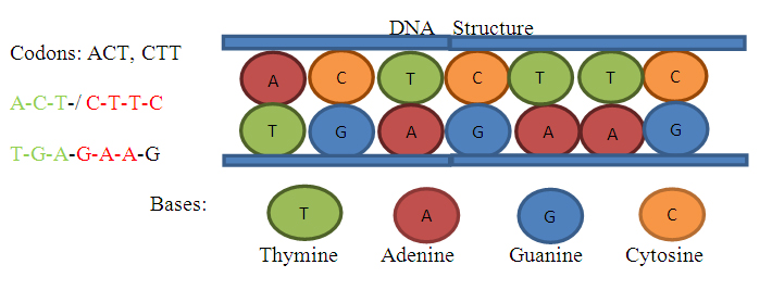
Figure 4 The DNA Molecule
The structure of DNA molecule, when unwound looks like a ladder. It is composed of two supporting "rails" made of sugar and phosphate molecules. The "rungs" of the ladder are the complementary base pairs. The base pairs are held together by hydrogen bonds. A sequence of three bases forms a codon, or gene.
Genes
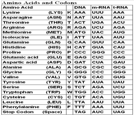
A gene is a section of the DNA molecule that is coded through its sequence for one specific protein. The human's genome has about 20,000 to 25,000 genes. 7 Our genes are coded with the information to make all the needed proteins that carry out the functions of the cells in our body. The sequence of the nucleotides, or base pairs, on each gene, determines the code to make a specific protein. When the code is translated or read, the sequence of nucleotides tells the ribosome to make a specific protein. Proteins are polymers of amino acids, which are coupled in sequence. One way to think of this is that the four bases, A, T, G, and C, are the letters in the DNA language. These letters are always bundled in groups of three, called codons or words. Each bundle, or word, ACT, CAT, GTA, AAT, represent a specific amino acid. For instance, the three nucleotides or codon, CAT, will specify the amino acid, histidine, when it is synthesized in the ribosome. Each word is read and then translated in the ribosome to make a sequence of amino acids. It is the sequence of the amino acids that build, or synthesize, a specific protein. In summary, a gene carries the code for an amino acid in its sequence of bundled bases called a codon. The genes in the DNA molecule go through a process of transcription and are then translated into the proteins they represent. In this way, the genetic information held on the structure of DNA is able to carry out the expression of its design.
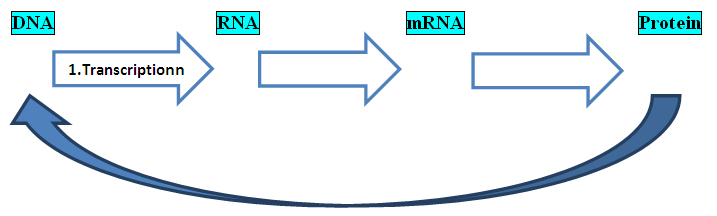
The Central Dogma of Biology
The central dogma of biology is the methodical process that the DNA molecule goes through in order to make a protein. It includes the three transitions. The DNA molecule goes through these three transitions in its expression of the encoded message held in the sequence of nucleotides, in this way the message on the DNA molecule is decoded and made into proteins. First, the message on the DNA, or gene, is transcribed or copied to RNA. Then the RNA undergoes processing. This processing involves splicing out non coded regions called introns before the completed messenger RNA (mRNA) molecule is made. Finally, translation of the mRNA occurs resulting in the synthesis of the proteins it is coded to make. 8 (see Figure 5) As a result, DNA is able to express itself through the proteins it ultimately makes.
 9
9
Figure 5 The Central Dogma of Biology
The central dogma is the series of processes that DNA goes through to make the proteins needed for the function of a cell. First, the DNA is transcribed or copied to RNA, then the non-coded areas are spliced out to make mRNA, or messenger RNA. The mRNA leaves the cell and the message it holds in its sequence of bases, or codons is translated into amino acids which build proteins. The proteins then help to build the DNA.
Transcription and Translation
Transcription is the process of copying the DNA molecule to RNA: a section of the DNA (deoxyribonucleic acid) transfers its code from the gene onto an RNA (ribonucleic acid) molecule. The transcription process starts when transcription factors mark the starting location of the gene. RNA polymerase recognizes the starting point and inserts itself between the double helix. Then, an activator protein initiates the process of transcription and the RNA polymerase speeds down the DNA strand, opening the double helix to expose the two strands. However, while the strand is opened, just one of the DNA strands is copied with complimentary bases resulting in the creation of a messenger RNA (mRNA) molecule. In transcription of DNA into RNA, the base adenine pairs with uracil instead of thymine. 10 When the DNA is transcribed, the exact strand is copied. This includes all the exons or coded areas and the introns, or non-coded areas. Next, the RNA molecule is spliced in the nucleus of the cell to remove the introns, or non-coded areas from the RNA.(see Figure 6) As a result, the new mRNA is pure uninterrupted code. The series of codons, or groups of codons are now ready to be translated into chains of amino acids or proteins. (polypeptide.) 11
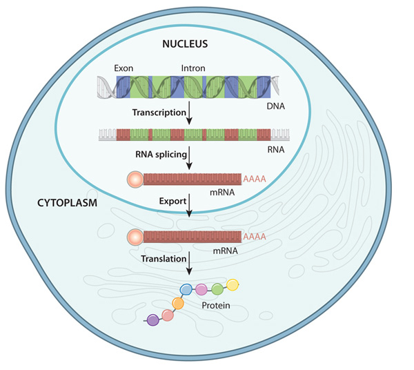
Figure 6 Processing of DNA into a Protein
DNA goes through a series of processes to make a protein. These processes take place in the nucleus and cytoplasm of the cell. Transcription is the process of DNA being copied to RNA. This process takes place inside the cell nucleus. The sequence of nucleotides on the DNA molecule is transferred to an RNA molecule while in the cell nucleus. The RNA molecule is spliced to remove introns, or non-coded areas. After the RNA is spliced, the mRNA moves out of the cell nucleus into the cytoplasm where it is translated into amino acids by the ribosome and then synthesized into proteins.
Translation
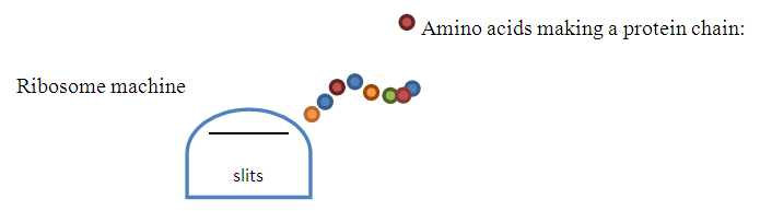
The messenger RNA (mRNA) holds the "message" of the DNA and travels out of the nucleus and into the cytoplasm.( see Figure 6) In the cytoplasm, the mRNA connects to an organelle called a ribosome. The ribosome acts like a protein factory, synthesizing proteins, which means it makes proteins. The ribosome translates each codon on the mRNA into an amino acid. Together these amino acids make a polypeptide chain or protein. This process begins when the mRNA travels through the ribosome "machine," and an amino acid is made from each bundle of three bases, or codon, forming a chain of amino acids. Codons are a group of three bases, or a "word" that name a specific amino acid to be made. (see Figure 7) This chain is made when the amino acids connect to each other after the ribosome reads each codon and translates it into the amino acid it names. The set of codons could also be thought of as the "word" for a specific protein. As the mRNA moves through the ribosomes, it translates the codons into the amino acids. As a result, each codon is translated into a specific amino acid and the message of the gene is expressed. 12
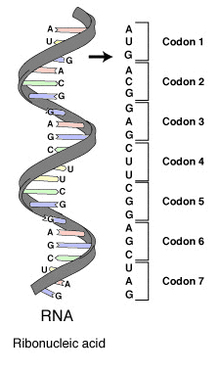
Figure 7 RNA Code for Amino Acids
The RNA molecule carries the codes that build the amino acids in the sequence of its bases. Each bundle of three bases forms a codon. Each codon will be translated at the ribosome for a specific amino acid. The sequence of the codons specify the sequence of amino acids in a polypeptide.( For example below codon 1: methionine (start), codon 2: threonine, codon 3: glutamate)
Proteins
Protein is a complex chemical made up of a chain of units called amino acids. Proteins are the " worker bees" of the cell. They perform thousands of functions necessary for the survival of the cell. A string or chain of amino acids is called a polypeptide chain. There are 20 natural amino acids. A protein can have hundreds of amino acids connected in specific order. Proteins can be made of one or more polypeptide chain. 13 The sequence of nucleotides on the mRNA will determine the proteins built. The various arrangements of the four bases on the mRNA are grouped into bundles of three bases called codons, each codon decoded for particular amino acids.( see Figure 7) There are 4 3 or 64 codon variations that translate into the 20 amino acids. 14 Therefore, an amino acid can be coded in more than one way. For instance: The amino acid, leucine, can be coded in four different ways, CUU, CUA,CUG, and CUC (see Appendix). After a protein is made, it is folded and then does its job. There are special codons that signal the beginning and ending of a sequence of amino acids. For instance, a polypeptide sequences would begin with the codon, AUG and ending codons could be either, UAA, UGA, or UAG. 15
DNA Replication
DNA replication is the process of making two exact copies of the genetic information on a double-stranded DNA, so that one copy can be given to each of the two daughter cells produced when a cell divides. 16 This process begins when the double helix is opened up and copied into two single-stranded DNA molecules called daughter strands. The double-stranded DNA is unzipped by the enzyme helicase at an incredible speed, 1,000 nucleotides every second. 17 Spinning the DNA as fast as a jet engine to open the two strands. 18 Then, the two open strands offer templates for each of the new double-stranded DNA strand to be built. DNA polymerase, an enzyme, functions as a catalyst to add the complimentary nucleotides onto the exposed single strand of DNA, which is now accessible on the strand. The polymerase needs a primer to begin this process. Primase sends a replication signals to the polymerase in the form of RNA primers, (RNA) primase, which initiates process. Polymerase synthesizes the new DNA by adding the complementary nucleotides to form the two new daughter strands, exact copies of the original double stranded DNA. When the helicase opens the double-stranded DNA into the two single strands of DNA, it looks like a fork. Consequently, the site where this process occurs is referred to as the replication fork. 19 20
Chromosomes
Figure 8 Chromosome
a. DNA is organized in bundles called chromosome. DNA is tightly packed in these bundles to fit in the nucleus of the cell. b. We have 46 chromosomes in our human genome, 22 autosomes from each parent and one sex chromosome. A karyotype is a picture of, the 23 chromosomes, the human genome. Each chromosome is numbered on the karyotype and organized by size.

DNA molecules are organized into tightly packed bundled called chromosomes. Our chromosomes are long pieces of DNA located within the nucleus of every cell.(see Figure 8a) Each cell has 23 pair of chromosomes or 46 chromosomes, which is what we call our human genome. We inherit 23 of our chromosomes (1 member of each pair) from each of our parents. As a result, every person has two of each gene, one from each parent. Twenty two chromosomes come from our biological mother and twenty two chromosomes from our biological father. These 22 chromosomes are called autosomes. The 23 rd chromosome is called the sex chromosome. This chromosome determines one's sex, XX for a girl or one X and one Y for a boy. When genes are studied or analyzed, a picture of the chromosomes numbered and arranged by size is often used. It is an excellent tool to study the human genome and chromosomal structures. This picture is called a karyotype. 21 (see Figure 8b)
Our human genome refers to the total DNA that comprises our 46 chromosomes. A common analogy of the human genome is, to think of the genetic code as a library, the chromosomes would be the books in the library, and the genes would be the chapters of each book containing directions for one particular function of the body. 22
Heredity
Heredity is the manner in which genetic traits are passed on to the next generation.
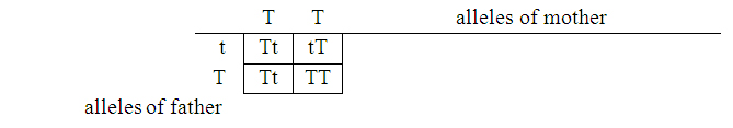
Chromosomes are the means through which the genetic material is transferred. The process of meiosis makes a sex cell or gametes, which holds just one set of a parent's chromosomes twenty-two plus a sex chromosome. During sexual reproduction, both sets of chromosomes are joined when the egg is fertilized by the sperm. Fertilization combines the genetic material from both parents, fusing to form a zygote, the first cell of a new individual. 23 As a result, the offspring has the combined genetic material of both parents. Consequently, each cell will have a pair of each chromosome, which results in a pair of genes for each trait. The positions of the two genes on the chromosome are alleles, or one's genotype. One allele is often dominant over the other. The dominant allele is expressed and can often be observed as physical trait, like eye color, right or left handedness, freckles, and curly hair. The observable trait is called one's phenotype, on the other hand, an unexpressed trait is called a recessive gene. We inherit many different traits including physical, behavioral, and predispositions to medical conditions like sickle-cell anemia, cystic fibrosis, heart disease, cancer, and some mental illnesses. 24
Genetic Disorders
Genetic illnesses are diseases caused by one or more damaged, mutated, or defective genes. These illnesses are extremely difficult to treat because the heart of the problem lies in the genetic code, which will continue to produce the condition unless it is corrected. Because the defective gene or genes for the illness is imbedded in the DNA, treatments have fallen short, only able to target the resulting effects of the disease produced by the ongoing construction of the destructive genetic codes. Finding, targeting, and correcting these genes is the focus, challenge, and hope of genetic engineering.
There are thousands of genetic illnesses: cancer, diabetes, asthma, melanoma, hemophilia, and sickle cell anemia are just a few. Some are genetic diseases are monogenetic, meaning they are caused by a single defective gene while other illness are polygenic, involving more than one defect gene. Other illnesses are caused because a whole chromosome or large segments of the chromosome are missing, duplicated, or damages as in Williams, Turners, and Downs Syndrome. Polygenic illnesses are more difficult to treat because more than one gene is involved. Monogenetic illnesses are cause by a single defective or mutated gene, which alters or eliminates the production of that gene's encoded protein. This one change can have catastrophic effects on human health as seen in diseases such as sickle -cell anemia, cystic fibrosis, Huntington's disease, and hemophilia. 25
Sickle Cell Anemia
Sickle cell anemia is an inherited blood disorder passed on through a single gene. This illness affects the ability of good hemoglobin (HbA) to carry oxygen. This genetic trait is recessive, and therefore can be carried genetically by a parent unnoticed. However, when a child inherits both damaged genes (one from the mother and one from the father), he acquires the illness. Sickle-cell anemia is a monogenetic illness, which means that the illness is cause by only one gene. This one problematic gene in located where the hemoglobin is coded. The defective gene, codon, which should be GAG, is incorrectly coded as GTG. 26 As a result, the ribosome reads GTG and makes the amino acid valine, instead of the correct amino acid glutamic, GAG. This one difference has a catastrophic effect on the ability of the hemoglobin molecule to carry oxygen. As a result, an abnormal form of hemoglobin (HbS) is produced, which is unable to carry the oxygen needed. Healthy red blood cells are flexible, disc-shaped cells that last about 4 month, are able to hold needed oxygen, and move easily through the blood vessels. The fragile sickle cells are sticky, stiff, and fragile resulting in moon-shaped blood cells that clot too easily, and break down after about 10-20 days (instead of the normal 120 days) causing anemia. Gene therapies are being explored to treat sickle cell anemia by replacing or altering the gene that causes it. 27
Cancer
Although there are many different kinds of cancers, these cells have some common characteristics. A cancer cell does not contribute to the function of the body. It does not differentiate itself into a muscle, nerve, or bone cell. Cancer cells have abnormally large nuclei and can have the wrong number of chromosomes from a normal cell. Most importantly, a cancer cell has the ability to replicate without limitation and has been called immortal. 28 An example of the cancer cell's ability to replicate could be noted in the historic story of Henrietta Lacks and the "Hela" cell. This cancerous cell was isolated without permission from a cervical tumor of a young woman named Henrietta Lacks in 1951. This cancer cell replicated prolifically in culture: scientists where having difficulty in finding cells that had this property. This cell was shared among scientists and continued to replicate, eventually contributing profoundly to the advancement of research and medical treatments.
Cancer has many genetic links. The mutated gene, p16 is thought to be associated with melanoma, the gene that codes for telomerase in turned off in normal cells, but active in cancer cells, the inherited gene, BRCA1 is proven to cause breast cancer. The prolific unrestrained replication of cells is a characteristic of most cancers and the advancement of technologies is linking genetic origins. Preventing and treating cancers is the focus of much research, development, and genetic therapy.
Gene Therapy
Gene therapy is the way DNA can be manipulate to treat illnesses caused by damaged, mutated, or missing genes. The treatment of genetic illnesses has been extremely difficult because the root of the problem has been beyond our reach, until now. We are at the dawn of a new era. In recent years, the fields of engineering, biology, and technology have joined, propelling the science of genetic engineering forward with increasing speed. This has advanced the study, treatment, and diagnosis of genetic illnesses. Genetic engineers are developing safe and efficient therapies to correct, alter or fix a gene and then return it to the cell where it will integrate itself into the existing DNA.
Gene therapy is the way an altered gene is used to treat a genetic disease but this is extremely challenging. The problematic gene must be located, a specific corrective therapy developed, a viable vector to deliver the gene, and then the gene needs to integrate into the cell's transcription and translation, ultimately altering the DNA permanent way.
Before one can develop a therapy, one must understand the nature and origin of the genetic illness. Genetic therapies for monogenetic illnesses target the one defective gene that cause cystic fibrosis, CF, muscular dystrophy, MD, hemophilia, and sickle cell anemia. Some gene therapies can introduce genes into a cell that are encoded with a sequence to produce therapeutic proteins like insulin. 29 Other therapies include replacing mutated genes with a healthy copy, silencing or inactivating the malfunctioning gene, and inserting a correcting gene, or a recombinant gene that will correct the problem or fight the disease. 30
Recombinant DNA technology refers to the recombining the DNA molecules in an environment outside the cell, then returning the recombined, or altered, DNA to a host cell where it will hopefully replicate and be transcribed and translated into protein. The recombination procedure is accomplished by cutting a piece of DNA—often a carrier DNA or vector with the enzyme called restriction endonuclease and adding a new section of DNA into the region of the cut. The new DNA might contain an altered or improved gene. The process is completed by rejoining the DNA with the linking enzyme, ligase, to "glue" it back together.
After an appropriate therapy is determined, one must decide on a vector. There are many kinds of vectors each with specific characteristic to consider when choosing one for a particular delivery. 31 A good vector is able to deliver the therapeutic DNA into the nucleus of a cell safely and effectively. In order to do this, the cells need to be targeted and the gene needs to be delivered into the nucleus where the DNA is located. Once the gene is in the nucleus, the new gene must be "activated" and integrated into the existing DNA of the host cell. This means that its code needs to be transcribed and translated into the new protein. Lastly, this "activation" must be without harmful side effects such as an immune response or a toxic reaction, which would render the therapy unusable. 32 Retroviruses are good vectors because of their ability to enter and integrate their genetic material into the host cell. They contain single stranded RNA as the viral genetic material and use the host cell's transcription and translation mechanisms to replicate itself. Reverse transcription makes two copies of the viral RNA forming a double stranded DNA molecule, which integrates into the host genome. In the end, the virus's encoded proteins are then transcribed and translated. 33
Silencing Genes: Small-Interfering RNA or siRNA
Diseases can be treated and cured by stopping the expression of a specific gene that is causing the illness. One therapy used to stop gene expression is the use of antisense therapies. Antisense therapies take a known gene sequence of a disease, and synthesize a strand of complementary nucleotides, or an oligonucleotide, which will bind to the mRNA of the targeted gene. When the antisense oligonucleotide binds to the mRNA, it is no longer able to translate its message. In this way, the gene is turned "off" unable to translate its genetic code into disease causing proteins. Therefore, a therapy can be developed for an illness by making an antisense oligonucleotide for the disease-causing gene. Although effective, the antisense oligonucleotides have a short life, which limits its use. 34
Cutting- edge research led by Mark Saltzman of Yale University has shown great promise in the use of siRNA to silence cancer-causing genes. This work has implemented revolutionary biodegradable vectors improving the safety and effectiveness of delivery. The siRNA is a double stranded, small interfering RNA molecule that silences the expression of the gene like the antisense oligonucleotide, but its double stranded structure is more stable than the single antisense oligonucleotides and can last for several weeks. Si RNA binds to the complementary bases of mRNA transcript. As a result, it disables transcription by "interfering" with mRNA. 35 In Saltzman's study, the isRNA was imbedded in biodegradable nanoparticle polymers to deliver the small-interfering RNA. The nanoparticles were densely loaded with siRNA and were able to silence the defective gene in a safe and effective way. The gene was silenced! This revolutionary work is offering great hope for the advancement of genetic therapies and changing the face of medicine as we know it . 36
Classroom Activities
Engineering is a science that is well suited for the classroom. It embodies high engagement, creativity, critical thinking, and cooperative problem solving.
The purpose of the classroom activities is to provide concrete experiences that reinforce abstract concepts. Most activities should be completed with a partner or in a group. This is important because it will provide needed opportunities for collaboration, cooperation, problem solving, and important discussions this forum will facilitate. The creation of a, "Science Wall" is a great way to display visual explanations of concepts by students; a pictorial display of collective learning. The "Science Wall" will also serve as a vehicle for reflection and discussion, and valuable support for vocabulary, concepts, and further inquiry.
Activity : DNA Model
Objective: Students will be able to construct a model of the DNA molecule in order to demonstrate an understanding of its structure. The model will show that DNA is made of the four nitrogen bases, adenine, thymine, guanine, and cytosine, which pair with their complementary base (A-T, C-G) and connect to the supporting rails of sugar phosphate molecules.
Students will build a model of a DNA molecule using gummy bears and twizzlers. The four different colored gummy bears will represent the four nitrogen bases. The twizzlers will represent the sugar carbon, phosphate backbones. Write a color code for the four nitrogen bases on the board that correspond to the four colors of gummy bears. Explain that adenine always pairs with thymine and cytosine always pairs with guanine. Then ask students to use the color code to make base pairs with the gummy bears. Students will use toothpicks to join the two gummy bears representing complementary bases such as; A-T and C-G. After students make each pairing, they will connect it to the rails forming what looks like a ladder. When the ladder is complete, have students twist it. The model will look like double-spiraled helix of DNA.
Activity : The Cell
Objective: Students will be able to illustrate and describe an analogy for the functions of the organelles within the cell. There are many possible analogies. A few examples are: a school, a city, a castle, a body, or a town.
After studying the structures and functions of the organelles within the cell, students will create posters for their own analogies. Students will create visual descriptions comparing a cell to an analogy of their choice. For example, a student may draw a school, naming and describing the parts compared to organelles and their functions. .Encourage students to use headings, captions, and text boxes to organized their and explain the comparative functions of the school part and each organelle. Students may illustrate, use cut outs from magazines, or both for this project. First, ask students to make a chart for their analogy; writing the name of each organelle in the first column, a description of the functions of each organelle in the second column, and the name of the corresponding part of the school (or their own analogy) with a comparison of the similar function to the organelle in the third column. The example of , "A Cell is Like a School" chart in this unit can be used as a guide. Students will use this charts as a resource to create a poster. Additional resources can be viewed online at: http://biology.unm.edu/ccouncil/Biology_124/Summaries/Cell.html
Activity: Observing DNA from Strawberries!
Objective: Student will be able to isolate, extract, and observe DNA from strawberries.
Materials: ziplock bags, strawberries, coffee filters, cold ethanol, extraction buffer ( detergent), 10mL pipette, 50 mL tubes,
Give each student a strawberry and a ziplock bag. Place the strawberry in the bag, seal, and mash. Measure 10ml of DNA extraction buffer with pipette and add to each bag. mash for 60 more seconds. Try to avoid making air bubbles. Fold coffee filter and fit into tube. Then carefully pour strawberry mixture into coffee filter until you have approximately 3mL of juice. Measure 10mL of cold ethanol with pipette and pour into mixture. You will see large bubbles and a white gooey mass. This is the DNA!! Remove the DNA. Have student write down the procedure and their observations. Take a closer look under a microscope. Can you see the strands when it is lifted? How did the DNA get out of the nucleus?
Activity: Singing About DNA
Objective: Students will work in groups to write songs about DNA that reflect knowledge of vocabulary, functions, and structure of DNA.
Divide class into groups of four or five. Ask students to work in groups to make a list of key words and phrases that reflect the functions and structure of DNA. Then join the class together and share lists. Write a collective list of DNA words and phrases from students' sharing. Post it. Next, ask students to think of familiar songs, note choruses, and verses. List them. Finally, set groups off with the challenge of writing a song that reflects what they know about DNA. Encourage students to use as many words from their DNA word list and to add movement to their songs for more fun. Celebrate with a Sing Off. Let your students go to other classes to share their songs spreading their enthusiasm for science! Extension: Make a Student Song Book. Have student type and illustrate their songs. Then copy and bind them into individual DNA Song Books.
Activity: Replication
Objective: Student will work in pairs to build a replication fork, simulating the process of replication.
Students will use connecting blocks to build replicated strands of DNA connecting complimentary pairs to each strand of the opened double helix.
Activity: Simulation of DNA Transcription and Translation
Materiel: red and yellow crate paper rolls, tables to decode codons to the protein they represent.
Objective: Students understand how the process of transcription and translation builds proteins from the DNA code through a simulation of the process.
Divide students into groups. Each group will draw the outline of a cell membrane and the cell nucleus on large paper. (3x3)
DNA: Making the DNA: Students can copy DNA code from the board or write their own onto a roll of red crate paper. You can write one of the base pairs and students will know to write its complementary base.
Transcription: Making a complementary RNA strand: Students will open the DNA, or cut the red DNA crate paper in half simulating the enzyme helicase opening the DNA. Student will then place yellow crate paper ( RNA) side by side to the red crate paper.( DNA-tape to hold it temporarily) to represent the RNA strand. Student will also have a cup of single bases, color coded, on little squares in a cup to use for matching base pairs. A,T,C,G. However, uracil will be matched with adenine on the RNA strand. Students will build the RNA strand by matching and gluing each complementary base of the DNA to the yellow crate paper.( a-u, c-g) Student will glue each color coded, labeled, complementary base pair onto the yellow crate paper roll one nucleotide on at a time.
Translation: First, student will move the messenger RNA out of the nucleus into the cytoplasm. Next, student will make ribosome machines(below)by cutting out a shape about 3" x 3" to represent the ribosome. Cut two slits in the shape which will allow the mRNA crate paper strand to be threaded through the ribosome. Then, as the students thread the mRNA strand through the ribosome, they will stop to view and translate each codon and use their protein reference sheet to decode each codon to the protein it represents. Finally, students will make colored circles to representing each amino acid built which they will connect to create a protein chain.
Activity: Proteins Bracelets
Objective: Students will understand that a protein is a sequence of amino acids that form a chain.
Assign a different colored bead to each of the 20 proteins. Ask student to make a codon pattern to for a bracelet. Students can use their reference guide (see Appendix) to transcribe each codon to the amino acid it represents. Students will then translate the amino acid into the assigned colored of bead. In this way, students will use two processes, transcription, and translation, to get a bead and then build a protein bracelet one amino acid at a time just like the ribosomes.
Activity- Inheritance Family Tree,Tracing Traits
Objective: Students will understand that we inherit our genome from our parents, and consequently all our observable genetic traits. These observable characteristics can be traced in our families.
Students will make a family tree that traces a line of inherited genetic traits.
Students will create a chart to collect data on observable genetic traits. Make a list of observable, inherited traits in the first column(curly hair, tongue roll, color blind, handedness, detached earlobes, freckles, ect.) Head the second column with the student's name, for a personal survey. In the third, fourth column ect. Collect data on your family members. Students will use this data to make a family tree that displays a path of inherited traits. Extentions: Chart class data for inherited traits.
Punnett Square: The Punnet Square can be used to show the dominant and recessive alleles for a specific trait. Students can use it to predict outcomes and probabilities of specific traits.
Activity- Genetic Engineering Simulation
Engineering design process to follow: Ask, imagine, plan, create, test, and improve
Group students for project: Students will pick a monogentic disorder to cure. First, students will study the disease to develop a comprehensive understanding of the illness. Next, a study of the diseases genetic characteristics will be explored, researcher and documented. This will include the location of the problematic gene and the protein it is producing. Students will use their engineering toolboxes, working collaboratively, to develop gene therapies that correct the mutated gene. Students will build stimulations to test solutions.
Bibliography
Nicholl, Desmond S. T. An Introduction to Genetic Engineering, 3 rd Ed. Cambridge: Cambridge University Press, 2008.
Watson, James D. The Double Helix. New York, New York: Simon & Schuster, 1996.
Mader, Sylvia. Human Biology. New York, New York: McGraw Hill, 2008.
Panno,Joseph. Ph.D. The Cell: Evolution of the First Organism. New York, New York: Facts On File, Inc. 2005.
Panno, Joseph. Ph.D. Gene Therapy, Treating Disease by Repairing Genes. New York, New York: Facts On File, Inc. 2005.
Saltzman, Mark W. Biomedical Engineering, Bridging Medicine and Technology. Cambridge, MA.2009
Saltzman, Mark w., Woodrow, Kim A. , Cu, Yen , Booth, Carmen J. , Saucier-Sawyer, Jennifer K. ,Wood, Monica J., Intravaginal gene silencing using biogradable polymer nanoparticles densely loaded with small-interfering RNA, Nature Materiels, Volume 8,, June 2009, Macmillan Publishers Limited, 2009.
Help Your Kids with Science, A Unique Step By Step Visual Guide. New York, New York: Doring Kindersley Limited, 2012
Websites Resources
http://www.dnalc.org/resources/3d/index.html
http://www.cellsalive.com/
http://www.genome.gov/10001177
http://ghr.nlm.nih.gov/handbook/therapy/genetherapy
http://www2.le.ac.uk/departments/genetics/vgec/highereducation/topics/dnageneschromosomes/dnagenechromoso
http://learn.genetics.utah.edu/
http://www2.le.ac.uk/departments/genetics/vgec/healthprof/topics/gene-therapy
http://www.genome.gov/Education/
http://www.dnalc.org/resources/3d/17-sickle-cell.html. Great animation for students of sickle cell anemia
http://kidshealth.org/teen/diseases_conditions/blood/sickle_cell_anemia.html
http://biology.unm.edu/ccouncil/Biology_124/Summaries/Cell.html
Appendix
Common Core Content Standards addressed in this unit:
The Common Core State Standards aim to increase depth of content knowledge by integrating subject matter across the curriculum. An additional objective of the Common Core Standards is to strengthen reading comprehension skills and student's ability to use and understand more complex text. This unit addresses these issues directly through relevant, engaging subject matter of bioengineering.
Scientific Inquiry: Scientific inquiry is a thoughtful and coordinated attempt to search out, describe, explain, and predict natural phenomena.
Scientific Literacy: Scientific literacy includes speaking, listening, presenting, interpreting, reading, and writing about science.

Notes
1. Sylvia Mader, Human Biology, p. 42.
2. Joseph Panno, Ph. D., The Cell: Evolution of the First Organism, p.40.
3. (http://www.cellsalive.com/cells/cell_model.htm)
4. http://www2.le.ac.uk/departments/genetics/vgec/highereducation/topics/dnageneschromosomes/dnagenechromoso
5. http://ghr.nlm.nih.gov/handbook/basics/dna
6. http://www.biology101.org/biologystudyguides/DNA.php
7. http://www2.le.ac.uk/departments/genetics/vgec/highereducation/topics/dnageneschromosomes/dnagenechromoso
8. Sylvia Mader, Human Biology, p. 444.
9. W. Mark Saltzman, Biomedical Engineering; Bridging Medicine and Technology, p.104.
10. Sylvia Mader, Human Biology, p. 449.
11. W. Mark Saltzman, Biomedical Engineering; Bridging Medicine and Technology, p. 102-104 .
12. W. Mark Saltzman, Biomedical Engineering; Bridging Medicine and Technology, p. 104-105 .
13. W. Mark Saltzman, Biomedical Engineering: Bridging Medicine and Technology, p.125.
14. W. Mark Saltzman, Biomedical Engineering; Bridging Medicine and Technology, p. 108 .
15. W. Mark Saltzman, Biomedical Engineering: Bridging Medicine and Technology, p. 137.
16. W. Mark Saltzman, Biomedical Engineering: Bridging Medicine and Technology, p. 98.
17. Joseph Panno, Ph. D.,The Cell: Evolution of the First Organism, p.66.
18. http://www.dnalc.org/resources/3d/03-mechanism-of-replication-basic.html
19. W. Mark Saltzman, Biomedical Engineering; Bridging Medicine and Technology, p. 98 .
20. Joseph Panno, Ph. D. The Cell: Evolution of the First Organism, p.64-65.
21. http://www2.le.ac.uk/departments/genetics/vgec/highereducation/topics/dnageneschromosomes/dnagenechromoso
22. http://genetics.emory.edu/docs/Emory_Human_Genetics_Chromosome_Analysis.PDF
23. Help Your Kids With Science, p. 43 .
24. http://learn.genetics.utah.edu/content/begin/traits/
25. Joseph Panno, Gene Therapy, p.1
26. Joseph Panno, Gene Therapy, p.1
27. http://kidshealth.org/teen/diseases_conditions/blood/sickle_cell_anemia.html
28. Sylvia Mader, Human Biology, p. 404.
29. W. Mark Saltzman, Biomedical Engineering: Bridging Medicine and Technology, p. 118.
30. http://genetics.emory.edu/docs/Emory_Human_Genetics_Chromosome_Analysis.PDF
31. http://learn.genetics.utah.edu/content/tech/genetherapy/gttools/
32. http://learn.genetics.utah.edu/content/tech/genetherapy/whatisgt/
33. W. Mark Saltzman, Biomedical Engineering: Bridging Medicine and Technology, p. 118.
34. W. Mark Saltzman, Biomedical Engineering: Bridging Medicine and Technology, p. 109.
35. W. Mark Saltzman, Biomedical Engineering: Bridging Medicine and Technology, p. 109.
36. W. Mark Saltzman, Kim A. Woodrow, Yen Cu , Carmen J. Booth, Jennifer K. Saucier-Sawyer, Monica J. Wood, Intravaginal gene silencing using biogradable polymer nanoparticles densely loaded with small-interfering RNA, Nature Materiels, p.526.
Comments (0)
-
 Public School Teachers Named Yale National Fellows
 -
 Yale National Fellow Featured on CBS 6 News in Richmond
 -
 Curricular Resources
 -

Search Curricular Resources written by teachers in National Seminars and Local Teachers Institute seminars.
-

View the Photo Gallery of Participants at Yale.
-

Explore the archive of News and Feature Stories.