An Advanced Explanation of the Electrical Impulse
The information that follows is a more specific and advanced explanation of the firing of an action potential. In this section, I have followed the formulation and utilized the circuit equivalents of cell membranes in the book Physical Biology of the Cell, Chapter 17, "Biological Electricity and the Hodgkin-Huxley Model."
Electrical Potential Creation and Use in Signal Creation
Cell membranes separate charge by the function of ion channels and ion-exchange pumps. Energy is consumed in this process, and stored in the form of electric potential: this is similar to our experience with batteries on the macroscopic level. The cells utilize this charge separation produced across their membrane by ion channels and pumps to keep the cell from returning to equilibrium. In addition to providing a necessary element for the survival of the cell, the cell membrane has the remarkable ability to propagate a signal along its length without the attenuation of the signal, in the form of an action potential. The voltage difference across the cell membrane is the physical basis for the creation of an action potential, which is the resulting disturbance caused by altering the polarity of membrane by a chemical, electrical or mechanical stimulus, which then propagates down the cell membrane (see Diagram 3). This mechanism is the fundamental basis for an animal to sense its environment and to communicate that information over a distance. In the broadest sense, it is the capacity for neurons to transmit a signal that enables the transfer and encoding of information that eventually results in the nearly inconceivable volume of information processing of the human brain.
Explanation of Biological Current
In the study of physics, we mostly discuss electricity as electrons flowing along conductors. Conventional current flow occurs in the positive direction, primarily because the original work done with batteries by Volta and Daniell in the late 1700s and early 1800s relied on the flow of positive ions such as Zn 2+, Cu 2+, and Ag +. In cells, there are a few cases in cells of electron transport directly producing charge separation, such as in photosynthesis in chloroplasts and electron transport in mitochondria. Usually, though, cells create and manipulate gradients of positive ions across the membrane. The most important ions in this process are Na +, K +, and Ca 2+. This makes sense because many of the large molecules in the cell, particularly nucleic acids, are intrinsically negatively charged and consequently, small positive ions are utilized in the creation of the cell's electrical state. Of the small negative ions, chlorine ions, Cl -, are the only monatomic ions exploited by cells processes. Bicarbonate, HCO 3 - , is another important anion in cell function.
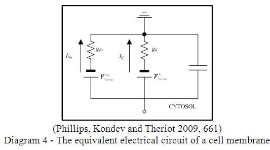
The electrical potential in cells is predominantly created by the nonequilibrium of sodium and potassium. The creation of electric potentials and currents requires cellular mechanisms that can separate and concentrate ions and control the flow of these ions through selected conduits. The cell membrane is a key component because its phospholipid bilayer contains many different ion-selective channels and pumps and is an effective thin insulator. The electrochemical properties of the cell membrane makes it equivalent to a circuit with a set of resistors (which are voltage-dependent), batteries (whose voltage is set by the ion concentration difference), and a capacitor which are all connected in parallel (Diagram 4). By selectively opening and closing the ion channels, which can only allow the transport of a specific ion, the cell can adjust its membrane electrical potential. This process is the key to electrical signaling in excitable cells. The neuron acts like a parallel RC circuit powered by the electric potential. We will create this model of a cell membrane in our classroom.
The Nernst Equation
The Nernst Equation enables us to quantify the electrical potential provided by the ionic gradient. In this case it is the cell membrane that we will consider. We can calculate the electrical energy mathematically by multiplying the ionic unit charge and the potential difference. We can also calculate the probability of finding an ion in a given region using the Boltzmann distribution for electrical energies. Although these formulas look complicated, setting up a proportion of concentrations allows us to take the logarithm of both sides of the equation and derive the famous Nernst equation,
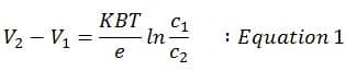
which will enable us to understand how cells utilize charge to create electric potentials. 3 We will consider this in more detail in the First Lesson Plan at the end of the unit where the students will learn how to use their knowledge of electrical physics equations and chemical ionic information to derive this powerful formula.
Speed of Propagation of Nerve Cells
The electrical signal propagation of neurons is a remarkable process that allows animals with nervous systems to respond to their environment fast enough to be able to survive. For animals, it is essential to be able to respond to stimuli in their environment rapidly, to avoid harm, elude predators, or catch prey. Chemical diffusion is a very slow process, as we know by the time it takes for perfume to diffuse to us in a room without drafts, and is insufficient for important signal conduction. The electrical conduction of a signal is an ingenious evolutionary adaptation. Action potentials transmit information 9-10 orders of magnitude faster than it would take molecules to diffuse over the length of a typical axon, and about 7 orders of magnitude more rapidly than motor-driven transport could send a signal the same distance. 4 The signal propagates through a single neuron at a rate of 10-100 m/s. The evolution of the action potential allowed for the survival of complex animals and eventually led to the development of the cortex (which is the outer, convoluted section of the brain responsible for higher level reasoning). The cortex enables the advanced critical thinking of the human brain.
Voltage-gated Ion Channels
Ion channels are proteins that allow the selective passage of ions through a membrane. When channels are voltage-gated that means that the state of the channel is dependent on the potential difference across the membrane. Voltage-gated ion channels are essential for the cell's control of the charge differential that enables the polarization of the membrane that is a signal spike, or action potential. Ion channels are able to detect stimuli such as a particular voltage change, permit the flow of select ions through the channel. The natural state of the ion channel is to be open, however, the negative normal potential that exists causes the protein of the ion channel to be closed. 5 Only when the threshold is reached, resulting in an action potential, the firing of which momentarily makes the membrane positive, do the voltage-gated ion channels open and depolarize the neighboring membrane, thereby propagating an electrical signal down the axon.
Membrane Depolarization and the Membrane as a Switch
The change of sign of the membrane potential is known as depolarization. The voltage-gated ion channels are essential as a switch for a signal to be created. The specialization of excitable cells to conduct an action potential occurs if the cells have selective ion channels that can be opened in response to a change in membrane voltage. When this happens, a local and temporary depolarization of the membrane can be amplified and propagated to travel across the entire length of the neuron. This propagated signal occurs quickly and without attenuation as indicated previously. These features are essential to the neurons ability to achieve its purpose.
The Electrical Circuit Model of the Membrane
The membrane can be idealized as an electric circuit. In terms of biological electricity, the system can be demonstrated as a collection of resistors, batteries, and capacitors, as shown in Diagram 4. The essential aspect is that the presence of the thin insulating membrane creates a capacitance to the system of membrane and channels, while the existence of ion channels makes the membrane behave like a series of resistors connected in parallel. The conductance of each channel type is different. The battery elements in Diagram 4 arise from the Nernst potentials that result from the concentration difference of ions across the cell membrane. The Nernst electrical potential for each ion can be represented as a battery because the voltage is additive and will have to be overcome or act in concert with the electrical potential across the membrane. 6
Mathematical Models of the Current
The difference between the potential of the membrane and the Nernst potential gives the driving force for ion movement or current: I=g(V m e m-V N e r n s t) Equation 2. The Nernst potential plays the role of a battery in series, which is additive, with a resistor of conductance g, as depicted in Figure 4. In this equation, a linear relation between the current and the voltage is assumed; this relation is known as Ohm's law. Ohm's Law as stated in physics is V=IR or I=V/R, which will be familiar to physics students.
In reality though, like much of the idealized physics equations, the voltage-gated channels actually result in a much more complicated, nonlinear current-voltage relationship across the cell membrane. The consequence of this is that when the potential of the cell changes so as to allow voltage dependent channel gating, the membrane permeability changes, due to the newly open channels. In the open state, the channels allow for current to flow in a fashion that is predicted by the I-V curve. A simple model for the ionic current through the membrane presented in Equation 2 above demonstrates a linear relationship between the membrane potential and the curve. Therefore, the ionic conductance, g, as defined above must be voltage-dependent. 7
Action Potentials and the Hodgkin-Huxley Model
Signals in cells are often mediated by the presence of electrical spikes called action potentials. At this point our interdisciplinary study enables us to quantitatively describe an action potential. The study of action potentials culminated in one of the most successful models for the integration of biology, chemistry, and physics in the history of science, the Hodgkin-Huxley (HH) model. The HH model depicts action potentials and predicts the nonlinear affect on the membrane model of alterations in the membrane voltage; it is a widely accepted explanation of neuron functioning. Through this model the action potential is described and its properties can be graphed.
The action potential is an all-or-nothing event. If a charge stimulus raises the membrane voltage above a threshold (V~-40mV-depending on the cell), the membrane potential jumps up to a value roughly given by the Nernst potential of sodium, V N e r n s t ( N a ) ~50mV (see Diagram 3). The drastic increase in electrical potential of a small section of the cell membrane causes the adjacent small section of membrane to go above the voltage-gated threshold, causing it to be depolarized. This effect then influences the next section of membrane to exceed its threshold, resulting in a wave of signal propagation known as an action potential. The HH model is the mathematical equation that governs the propagation of an action potential and it provides an indication of how electrical signals can be propagated over long distances at constant speed and without dissipation. One other consequence of the electrical stimulation exceeding the voltage-gated threshold is that the membrane is temporarily desensitized and is incapable of further depolarization. This effectively allows the signal to only propagate in one direction, which is essential for the proper functioning of a neuron. 8
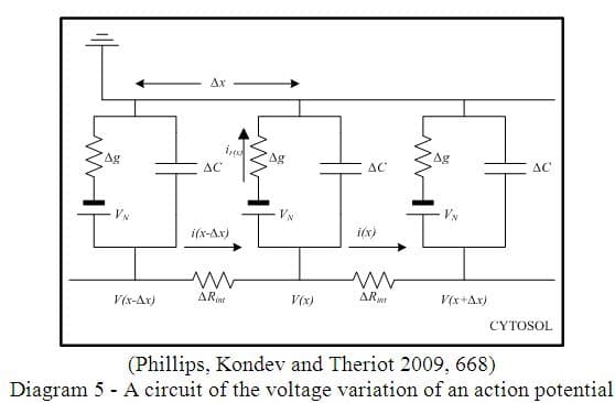
We can utilize the diagram above (Diagram 5) to simulate the action potential voltage variability over distance. Cell membranes and their ion channels can be modeled as a collection of resistors, capacitors, and batteries. Excitable membranes behave as bistable switches. The interaction between voltage-gated potassium and sodium ion channels mediates the response. The Hodgkin-Huxley model of the propagation of action potentials shows how cells can produce propagating pulses, or action potential spikes, that serve as the key information carrier in complex organisms. 9 This is meant to demonstrate the usefulness of the Hodgkin-Huxley model, to explain the electrical aspects of the neuron in physical terms, and to suggest lab possibilities and potential mathematical and graphical problems. I believe this physical knowledge justifies the pursuit of the HH model in the explanation of vagus nerve stimulation treatment, which is related to my patent and helps to illuminate the potential reasons for the onset of epileptic seizures.
Comments: