Overview of the Circulatory System
Blood supplies every cell in the body with oxygen and nutrients and removes waste products that could harm those cells. The heart circulates blood via a system of connected tubes called arteries, capillaries, and veins, which distributes the blood throughout the body. The following sections rely heavily on the text of Medical Physiology by Boron and Boulpaep, 2 although the information is of a general nature that should be consistent with any biology text that deals with the circulatory system. Similar material is also available on internet websites. 3
Overview of the Heart
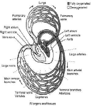
Figure 1 Overall structure of the circulatory system
Courtesy of W. Mark Saltzman 4
The heart is a set of muscular compartments whose contractions pump blood and a set of one way valves that ensure that blood flows in only one direction. The heart is divided into a right section and a left section and both sides are divided into an upper chamber called the atrium and a lower chamber called the ventricle. The right side of the heart is responsible for the pulmonary circulation between the heart and the lung, while the left side is responsible for the systemic circulation between the heart and the rest of the body. The time period when a chamber of the heart is relaxed is called diastole, and the period when a chamber of the heart contracts is called systole.
In a typical cardiac cycle, deoxygenated blood fills the right atrium from the inferior and superior vena cavae, two large veins that return blood from the body to the heart. Blood begins to flow directly from the right atrium into right ventricle through the tricuspid valve even before the atrium contracts. An electrical signal originating in the right atrium at the sinoatrial node causes the atrium to contract (atrial systole). The contraction forces the remaining blood in the atrium into the right ventricle. As the right ventricle fills with blood, the electrical signal from the heart arrives at the ventricle, causing it to contract (ventricular systole). The ventricular contraction increases pressure in the blood within the chamber, which causes the pulmonary valve to open, and forces blood out of the heart and into the pulmonary artery sending blood to both lungs. Under pressure from the ventricular contraction, the tricuspid valve closes, preventing blood from flowing backwards into the atrium.
Within the lungs, blood absorbs oxygen and releases carbon dioxide. Blood flow from the lungs returns through the pulmonary veins to the left atrium of the heart. The newly oxygenated blood flows into the left atrium and begins to flow through the mitral valve directly into the left ventricle before the atrium contracts. The left atrium contracts (atrial systole), sending the remaining blood into the left ventricle. After the left ventricle fills, it contracts (ventricular systole), pressurizing the blood and pumping it through the aortic valve and out the aorta and into the rest of the body. During ventricular contraction, pressure in the ventricle closes the mitral valve, preventing backflow into the left atrium.
While the process I described above makes it seem like there is a series of four consecutive contractions, in point of fact, both atria contract nearly simultaneously and later both ventricles contract nearly simultaneously. The right side pumps deoxygenated blood to the lungs, while the left side receives and pumps out oxygenated blood coming from the lungs from a previous cardiac cycle.
Overview of Blood Vessels
Oxygen-rich blood—pressurized for transport by the left ventricle—travels to each cell in the body, flowing through a series of blood vessels called arteries that branch (or bifurcate) into smaller vessels called arterioles, which further bifurcate into even smaller vessels called capillaries. The capillaries have extremely thin walls that allow the red blood cells in them to interact with the body's cells. While in capillaries, the red cells release oxygen and nutrients to the somatic cells and simultaneously pick up waste products and carbon dioxide. The blood then begins its trip back to the heart. The capillaries merge into larger vessels called venules, and the venules merge into larger veins. All of the veins merge into the inferior and superior vena cavae that lead back to the heart.
The arterial side of the circulatory system is under pressure, and it is this side of the circulatory system where blood pressure is taken. In the process of bifurcation, the diameter of the vessels decreases exponentially as the number of blood vessels grows exponentially to spread the blood over a greater volume. As the vessels get smaller, both the pressure in them and the velocity of the blood through them decreases. The slow velocity of the blood in the capillaries increases the time for exchange of molecules between the red blood cells and the somatic cells of the body.
When the doctor reads my blood pressure as 130 over 85 (135/85), the first number is the peak diastolic pressure generated by the left ventricle when it contracts. The second number is the systolic pressure, which is the residual pressure that remains in the artery when the left ventricle relaxes. I'll discuss the units of the numbers in these measurements shortly.
The venous side of the circulatory system is not under pressure. Incidental and natural movement of the skeletal muscles squeezes blood through the veins, and a system of valves within the veins prevent back flow.
Comments: